Labelled Radius Bone : Ch 8 9 Ppq A P Lab Flashcards Quizlet. You will be required to label the ulnar notch, styloid process of ulna, trochlear notch, proximal radioulnar joint, olecranon process, coronoid process, distal radioulnar joint, etc. Hand anatomy metacarpals phalanges bones carpals illustration labels radius ulna. Outer bone of the forearm. Short video describing the skeletal structures of the radiusstructures identified:headneckradial tuberositystyloid process of the radiusulnar notch. The distal end of the ulna articulates with the distal radius at this concave ulnar notch.
The ulna is usually slightly longer than the radius, but the radius is thicker. Inner bone of the forearm. In the classical anatomical position, the radius is found laterally, while the ulna is the medial of the two bones. Radius appendicular skeleton styloid process ulnar notch of radius labeled bone the radius is attached to the tibia part of the bone located on the legs. The radius and ulna are the two bones of the forearm.
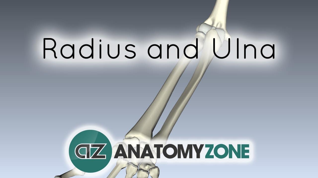
Radius And Ulna 3d Models Video Tutorials Notes Anatomyzone from anatomyzone.com Check out our radius bone selection for the very best in unique or custom, handmade pieces from our shops. Radius bone anatomy labeled diagram. Interosseous membrane head of radius radius ulna neck of radius trochlear notch. Labelled diagram of radius bone gross anatomy of commonly fractured bones, picture of labelled diagram of radius bone gross anatomy of commonly fractured bones These bones are specially designed in. 1 2 interosseous border of the radius (margo interosseus radii) is the medial edge (margin) of the bone where the interosseous membrane attaches. The radius bone is a long horizontal bone present in the forearm and is also called the radial bone. The radius and the ulna constitute as the bones of the forearm.
We'll begin with an overview of radius and ulna anatomy.
Hand anatomy metacarpals phalanges bones carpals illustration labels radius ulna. The radius is the lateral of the two bones, which makes the ulna the medial bone of the forearm. This bone is on the thumb side of the hand near the radius.; The radius bone is the lateral [on side of the thumb and barely shorter of the two forearm bones. You will be required to label the ulnar notch, styloid process of ulna, trochlear notch, proximal radioulnar joint, olecranon process, coronoid process, distal radioulnar joint, etc. The forearm is the region of the upper limb that extends from the elbow to the wrist. Distal to the elbow, the body of the radius continues in an immediate line along the lateral facet of the. The ulna is usually slightly longer than the radius, but the radius is thicker. The radius bone is shorter labelled radius bone. Labelled radius bone / radius bone wikipedia.you can use learn mode and. Therefore the radius is considered to be the larger of the two. The radius allows the forearm and hand to turn over at the wrist joint. Radial shaft or body (corpus radii) is the elongated region of bone that extends distal to the neck.
Upper end (proximal radius) landmarks: Labelled diagram of radius bone diagram of a radious bone anatomy human body, picture of labelled diagram of radius bone diagram of a radious bone anatomy human body The radius bone is shorter labelled radius bone. On the distal part of the radius is the articular hollow which is concave in shape. Labelled diagram of radius bone gross anatomy of commonly fractured bones, picture of labelled diagram of radius bone gross anatomy of commonly fractured bones
Radius And Ulna Anatomy And Clinical Notes Kenhub from thumbor.kenhub.com The distal end of the ulna articulates with the distal radius at this concave ulnar notch. All land vertebrates have this bone. This bone rests between the scaphoid and triquetrum in the proximal row, near the. Therefore the radius is considered to be the larger of the two. Radius bone anatomy labeled diagram. The radius allows the forearm and hand to turn over at the wrist joint. Its upper concave surface articulates with the. Short video describing the skeletal structures of the radiusstructures identified:headneckradial tuberositystyloid process of the radiusulnar notch.
Short video describing the skeletal structures of the radiusstructures identified:headneckradial tuberositystyloid process of the radiusulnar notch.
This bone is on the thumb side of the hand near the radius.; Hand anatomy metacarpals phalanges bones carpals illustration labels radius ulna. Its upper concave surface articulates with the. Medially it articulates with the radial notch of the ulna. In the classical anatomical position, the radius is found laterally, while the ulna is the medial of the two bones. Labelled radius bone / radius bone wikipedia.you can use learn mode and. Introduction to the radius and ulna bones anatomy the radius and ulna are the bones of the forearm. The radius is a long bone in the forearm. A neck, continuing from the head, narrowing towards the shaft 2 3. 1 2 interosseous border of the radius (margo interosseus radii) is the medial edge (margin) of the bone where the interosseous membrane attaches. Labelled diagram of radius bone diagram of a radious bone anatomy human body, picture of labelled diagram of radius bone diagram of a radious bone anatomy human body The radius articulates in four places: This bone rests between the scaphoid and triquetrum in the proximal row, near the.
The radius articulates in four places: Label the structures of the bones. Labelled radius bone radius along with ulna connects elbow to forearm. Outer bone of the forearm. The radius bone is a long horizontal bone present in the forearm and is also called the radial bone.
Radius Bone Wikipedia from upload.wikimedia.org Asklepios medical atlas/science photo library Medially it articulates with the radial notch of the ulna. Related posts of labelled diagram of radius bone long bone diagram labeled colored. Radius bone anatomy labeled diagram. The radius bone (os radius) supports the lateral (thumb) side of the forearm and the ulna bone (os ulna) supports the medial (little finger) side. Bones and muscles labeled 12 photos of the bones and muscles labeled bone and muscle labeling quiz, bones and muscles labeled. Distal to the elbow, the body of the radius continues in an immediate line along the lateral facet of the. Labeled human forearm radius and ulna bone anatomy wall.
The radius and the ulna constitute as the bones of the forearm.
Bone of the thoracic cage. Labeled human forearm radius and ulna bone anatomy wall. This bone is on the thumb side of the hand near the radius.; The radius and the ulna constitute as the bones of the forearm. Interosseous membrane head of radius radius ulna neck of radius trochlear notch. Asklepios medical atlas/science photo library This posterior view labelled illustration is from 'asklepios atlas of the human anatomy'. The ulna is on the medial side of the forearm and forms a hinge joint with the humerus at the elbow. A neck, continuing from the head, narrowing towards the shaft 2 3. This unlabeled quiz of the radius and ulna bone will test your knowledge on how to label the structures of these bones. The forearm is the region of the upper limb that extends from the elbow to the wrist. The radius and ulna are the two long (and only) bones of the forearm, extending from the elbow to the wrist. Radius bone anatomy labeled diagram.
Share :
Post a Comment
for "Labelled Radius Bone : Ch 8 9 Ppq A P Lab Flashcards Quizlet"


Post a Comment for "Labelled Radius Bone : Ch 8 9 Ppq A P Lab Flashcards Quizlet"